Blending two technologies
March 02, 2016 | Wednesday | Views | By BioSpectrum Bureau
Blending two technologies
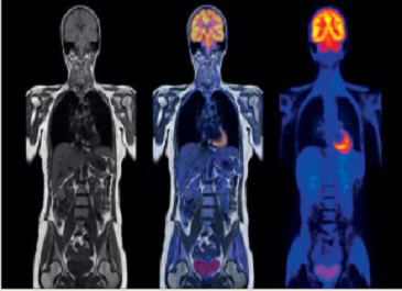
An MRI (left) shows even the smallest changes to tissue and organs, while a PET scan (right) shows the activity in specific parts of body. A combined molecular MR image (center) gives additional information for a more comprehensive assessment of the disea
MRI is a long-established and ever-improving technique for imaging structures in the body. On the other hand, Position Emission Tomography (PET) is used to show metabolic processes in the body. In the past, the two techniques were used sequentially if MRI alone was not sufficient for a diagnosis. However today, both MRI and PET are united in a single scanner - the molecular magnetic resonance (mMR) system.
How does mMR work?
MMR is an innovative hybrid imaging technology that captures metabolic, functional and structural processes at the same time. This new technology makes a single reference image by combining both techniques -- a tremendous advancement in diagnostic accuracy and precision.
Magnetic resonance imaging is based on the physical principle of nuclear resonance. It uses strong magnetic fields and alternating electromagnetic fields to excite the hydrogen nuclei in the body. As the nuclei return to a rest state they give off electrical signals, which the MRI scanner detects and uses to construct medical images. The results are precise 3-dimensional images of tissue structures.
With the simultaneous acquisition of MR and PET data, this system is designed to provide new opportunities for imaging. While MRI exams show the structural and functional detail of the human body, a PET scan shows the body's metabolic processes - its functions. The images are acquired by injecting a very small dose of a radioactive substance that travels through the body and is absorbed by the organs and tissues. The PET imaging device detects and records the energy given off by the tracer substance, thus making metabolic and biochemical processes in the human body visible. The results are usually 3-dimensional color-coded images of biological processes.
The innovative system has the potential to be a particularly valuable tool for identifying neurological, oncological and cardiac conditions of disease and in supporting the planning of appropriate therapies The mMR also opens new opportunities for research, such as the development of new biomarkers or new therapeutic approaches. One of the biggest advantages of simultaneous MR and PET imaging is reduction in examination time by almost 50%.
Applications
Neurology: Bringing MR and PET together offers the potential for a more complete imaging picture and better understanding of neurologic pathologies. With new tracer development, the simultaneous MR-PET system may add to the capabilities of imaging in general for neurodegenerative diseases or psychological disorders.
Oncology: In oncology imaging you can make full use of the unique features offered by synergetic MR and PET imaging. It helps deliver not only important information during early detection and staging, but also becomes a decisive factor for treatment planning, therapy selection and monitoring as well as follow-ups.
Cardiology: Comprehensive 2D routine cardiac applications, ranging from morphology and ventricular function to tissue characterization are possible, also enabling valuable diagnostic information about even subtle changes in tissue composition.
Paediatrics: Tissue relaxation times in pediatrics are very different compared to those of adults. The reasons for these differences are: developing tissues, body size, faster heart rates and compliance with breathhold commands. Especially in pediatric oncology, mMR adds an additional value as no ionizing radiation from MRI leads to significantly improved overall dose.
The Biograph mMR exam process
Patient is injected with the radiotracer fluid necessary for the mMR examination. During the rest period that follows, the fluid will go to various parts of the body through normal metabolic process. During the scan, the patient lies comfortably on a table. The body areas to be scanned are covered by so-called "coils" and the table slides into the machine's bore where the images are acquired.
For the first time, two patient-friendly imaging procedures are together in a single scanner: magnetic resonance imaging (MRI) to visualize the structural and functional detail of organs, and positron emission tomography (PET) tocapture the metabolic activities in the human body.
Where sequential imaging procedures were required in the past, Biograph mMR now has the power to perform a single,simultaneous scan and to obtain acomprehensive diagnostic picture. Not only does this result in shorter scan times, it can also lead to faster diagnoses and improved diagnostic confidence.
A technical revolution
Until now, it was nearly impossible to integrate MR and PET technologies: the conventional PET detectors, which use photomultiplier tubes, could not be used in the strong magnetic field generated by an MR system. Integration was further limited by the lack of space inside the MR device. For this reason, MR-PET imaging was the result of two separate scans (MR and PET) with a significant time lag. With Biograph mMR, Siemens brings the first molecular MR system for clinical research that integrates MR with compact, specialized PET detectors. The Biograph mMR - incorporating Tim, the "Total imaging matrix" technology from Siemens may make it even quicker and easier for clinicians to perform the MR examination.
Advantages of an mMR system
Siemens was the first in the world to introduce such a system called Biograph mMR
• Simultaneous acquisition of morphology,function and metabolism of the human body
• High diagnostic confidence
• Patient-friendly exams, reduced radiation exposure
• Shorter exam times
• Suitable for children, especially for metabolic anomalies.