Siemens Healthcare introduces first Twin Robotic X-Ray system
October 29, 2015 | Thursday | News | By BioSpectrum Bureau
Siemens Healthcare introduces first Twin Robotic X-Ray system
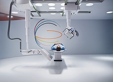
The two ceiling-mounted arms on Multitom Rax can be moved into position automatically using robotic technology (Photo courtesy: Siemens Healthcare)
Siemens Healthcare launches the first Twin Robotic X-Ray system, Multitom Rax (Robotic Advanced X-ray) at the University Hospital Erlangen today.
Multitom Rax (Robotic Advanced X-ray) enables a wide variety of examinations in a range of clinical areas to be performed using only a single X-ray system for the first time. In addition to conventional 2D X-rays, the system also makes it possible to perform fluoroscopy examinations, angiography applications and even 3D imaging. The operator is always in full control of the system's movement. By the push of a button, both robotic arms are being positioned fully automatically around the patient, improving both safety and convenience. There is no need to move the patient on the system or to change rooms for further imaging procedures, which makes examinations less painful and less time-consuming. Work processes in hospitals can be improved and economic efficiency increased.
"We see the Multitom Rax as a universal device that covers all aspects of X-ray diagnostics. You could call it radiology's answer to the Swiss army knife," says Prof. Michael Lell, senior physician at the Imaging Science Institute of the University Hospital Erlangen.
The new system can be used in a wide range of applications, from emergency medicine to orthopedics, angiography or fluoroscopy, and can thus help optimize clinical work processes. The fact that the detector can be freely positioned means that quite different X-ray images, both static and dynamic, can be taken in a single room using a single system. That saves time and avoids unnecessary costs, since specially installed modalities for examinations that are not performed on a daily basis can be uneconomical for hospitals. On the other hand, systems that are in regular use can cause lengthy waiting times, and this is where the new X-ray scanner can help ease the burden. Multitom Rax makes work processes economically efficient, while still being able to offer a wide range of examinations.
The two ceiling-mounted arms on Multitom Rax can be moved into position automatically using robotic technology, and they can also be moved manually, servo motor supported, when required - to make fine adjustments, for example. While one arm moves the X-ray tube and the large touchscreen, the other carries the 43 x 43 cm flat panel detector, which can record static, dynamic and real 3D sequences. "The robotic technology ensures a new level of precision and automation, enabling a new level of standardization and throughput", explains Mr Francois Nolte, head of the X-ray Products Business Line at Siemens Healthcare. He added, "The precise positioning of the arms in all three planes makes the examinations so much easier: regardless of whether the patient is standing, sitting or lying down, the robotic arms move with perfect accuracy using robotic technology. Our strategy is based on the principle that the system moves, not the patient, which reduces risk of additional injuries and pain."
With conventional radiography systems, the detector often has to be placed in an external holder. In addition to the extra time required, this also involves the challenge of positioning the tube at exactly 90 degrees. Multitom Rax does this at the push of a button for free exams. This also prevents any risk of having to repeat image processes because the tube was not precisely positioned. The system offers optionally also wireless, portable detectors in two different sizes that can be positioned directly between the wheelchair or mattress and the patient's back, which avoids the need to sit the patient up. The automatic control of the robotic arms ensures that they will always take the shortest and safest route to reach the next programmed position. Pre-programmed safety zones and an automatic stop in response to contact also improve safety.
3D computed tomography (CT) images are often used in situations such as orthopedic examinations involving the implantation of prosthetic joints, for example, to ensure that the artificial joint is best adapted to fit the patient's anatomy. Now, for the first time, Multitom Rax makes it possible to take 3D images under the patient's natural weight bearing condition. 3D images can be made of all areas of the body with the patient seated, lying down or standing. Images taken while the patient is standing are essential because for example knees, pelvis and spinal column appear differently under the influence of the patient's body weight compared to when the patient is lying down.
As a result, 3D images acquired by Multitom Rax offer better diagnostic and planning certainty compared to those that do not reflect a natural weight bearing condition. Conventional 2D x-rays, for example, do not always reveal fine hairline fractures in the bone. If a bone fracture is suspected, it has previously been necessary to take a 3D image using a CT system to be sure of the diagnosis. With Multitom Rax, however, a 3D image can be taken at the same system, and so the patient does not have to wait for a further appointment or to be transferred to the CT unit.
A free-standing patient table and fully mobile system elements with Multitom Rax provide a more comfortable examination atmosphere. The system is designed for all patient types, from children to the elderly, mobile, immobile and adipose individuals. The fact that the table can be adjusted to a very low 50 centimeter table height means that children can get onto it by themselves. It can also be positioned at the most convenient working height. The hospital staff thus has full access to the patient, with no need for the hospital staff to twist into an anatomically unnatural position. The result is an improvement in both safety for the patient and the examining physician, and in the level of convenience, since it is the system that moves when needed, not the individuals. Additional devices and personnel are often essential for interventional procedures such as fluoroscopic needle localization in particular. The open system design makes it possible to position the tube and detector most appropriately in the room. And the fact that both arms are ceiling-mounted means there is no floor-mounted equipment or cable ducts to get in the way.
Care (Combined Applications to Reduce Exposure) applications support treatment standardization using Multitom Rax and aim to keep the radiation dose as low as possible for both patients and hospital staff. Removable scatter grids and a copper filter, combined with the sensitive detector, help to minimize dose. Precisely focusing on the area of the body to be X-rayed and avoiding the need to repeat examinations using X-rays helps protect patients from unnecessary exposure to radiation. In the case of fluoroscopy examinations, such as gastrointestinal or swallow exams, several Care features keep the dose low. A preliminary examination using an especially low radiation dose to fine-tune the tube and detector helps to correctly position even in very challenging exams. For all examinations, in addition, the dose used is automatically reviewed and recorded.
As a part of the Max system family from Siemens Healthcare, Multitom Rax stands out by providing the same image impression and thus making it easier to compare X-ray images. The controls and user interfaces on the Max systems are identical, which means the operators have no need to familiarize themselves over again with new equipment. The wireless detectors in the Max family can also be used equally with all the systems in the family, improving the level of flexibility.
Multitom Rax is also configured to accommodate future trends in treatment with functions that can be adapted at a later time. And lastly, its closed surfaces are easy to keep clean, which contributes to the long service life of the system


