Researchers design the world's first 3D color X-rays
July 19, 2018 | Thursday | News
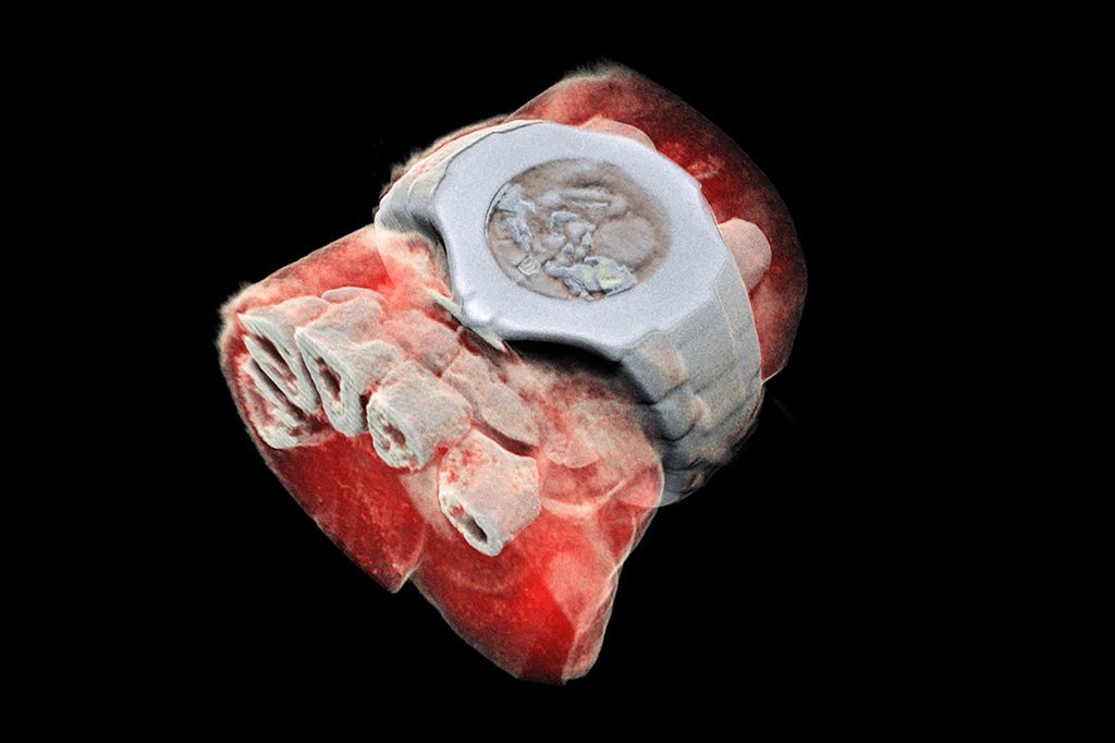
A new medical scanner, derived from technology used by particle physics researchers at CERN which can provide 3D colored X-rays
Scientists in New Zealand have designed an x-ray scanner capable of capturing human bodies in full color and three dimensions, which could give doctors a clearer picture for diagnosing cancer and other diseases, minimizing the need for invasive surgeries.
The new scanner has its origins in a tool that contributed to research into the universe’s fundamental particles and functions much like a camera. It counts subatomic particles as they meet pixels when its electronic shutter is open. That allows it to generate high-resolution images of soft tissues, including minute disease markers.
“We can make out details of various tissues, like bones, fats, water and cartilage, all functioning together inside the human system,” said Anthony Butler, a radiologist at Otago University in New Zealand, who developed the scanner with his father, Phil Butler, a physicist.
Whereas traditional scanners send x-rays through the body and show only two colors, white where bone tissue has absorbed the beams, and black where soft tissues have not, the new machine is sensitive enough to detect specific types of tissue (such as bone, cartilage, fat, and water) by analyzing individual light particles.
The new scanner matches individual X-ray photon wavelengths to specific materials, such as calcium. It then assigns a corresponding color to the scanned objects. The tool then translates the data into a three-dimensional image.
The tissue data are then used to construct a full color reconstruction of the body. The scanning technology was adapted from a tool that physicists used at CERN, the European particle physics laboratory near Geneva, Switzerland, to detect particles moving through the Large Hadron Collider.