Clinical Value of Ultrasound in Urology
June 16, 2020 | Tuesday | News
Emerging technologies in ultrasound offer optimal support for urology precision
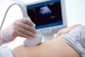
image credits: depositphoto
Ultrasound modality has grown leaps and bounds in India to become an inevitable part of Healthcare delivery. Urology is one of the applications where Ultrasound delivers tremendous clinical value as high-level of accuracy and reliability is necessary to ensure early detection, precise diagnosis and appropriate treatment. Emerging technologies in ultrasound offer optimal support for urology precision. High resolution B mode, Doppler mode, advanced imaging technologies such as Real-time Virtual Sonography, Real-Time Tissue Elastography and Contrast enhanced ultrasound lead to safer, more accurate diagnosis and treatment. A diverse selection of Probes provides versatility for different procedures.
Superior Technologies for Urology
Real-time Tissue Elastography is a technology where applying a very small, repetitive compression to the target with the ultrasound probe induces tissue strain which is displayed as a color map that represents tissue stiffness. It has proven clinical value for routine use across a variety of different applications including Prostate imaging. Simultaneous Dual Display of Contrast enhanced with fundamental B-mode image enhances display of microvasculature,
providing extra criteria for more precise characterization of lesion, facilitates understanding of location and extent of tumours and allows for prostate gland and kidney examinations. Inflow Time Mapping is a colour scale used to code the time-to-peak enhancement of the contrast agent and allows better differentiation of tumours by comparing the inflow time arrival pattern
between the lesion and the healthy parenchyma of the prostate gland.
Real-time Virtual Sonography (RVS) is an image guidance technology for interventional procedures. It enables direct comparison of lesions by taking advantage of the strengths of each imaging modality (Ultrasound & MRI) and targeted biopsy can be performed on lesions detected by MRI. RVS allows for Multi-modality imaging which is the combined use with Contrast Enhanced Ultrasound, Real-time Tissue Elastography etc.
Versatile Probes for Urology applications
Real-time bi-plane Probes simultaneously display sagittal and axial images of the prostate gland, providing a better understanding of the anatomical lesion location and allowing accurate biopsy targeting with needles, visible in both cross sections. End-fire Probes offer a FOV of 200 degrees and free choice of imaging planes for diagnostic and trans rectal biopsy. Side-fire bi-plane
Probes are used for trans perineal biopsy and grid template guided biopsies for brachytherapy treatment planning. 360°electronic radial trans rectal probe is used to assess rectal cancer staging with less patient discomfort. Dedicated Centre Biopsy Convex probes are useful for PCNL/Renal ablations or trans perineal biopsies for patients with rectum resection. A range of High and mid frequency linear probes provide high quality image performance for scrotum & testicular assessment to check anatomy & symmetry, vascularity and abnormalities.
Innovations in Ultrasound-guided Surgery
The Robotically measured ultrasound probe is a significant add-on that permits Surgical Robots to be used for multifarious procedures like Nephrectomy surgery where the Real-time imaging directly provides valuable information essential to help in surgical execution and planning Robotically controlled Ultrasound Probe, connected to the Surgical Robot, provides surgeons with direct control of the real-time imaging during minimally invasive RAPN procedures. After engaging the probe with the robotic graspers, the surgeon can completely control the probe’s movements from the robotic consoles rather than directing a surgical assistant.
4-way flexible linear probe for laparoscopic surgery can be inserted through a trocar for llocalizing the extent of tumour, confirming the presence of surrounding vessels, and detecting other lesions. Trapezoid imaging maximizes the view angles with Harmonic Imaging & Real- Time Tissue Electrography modes. 2-way & Rigid Laparoscopic Probes provide instant feedback
on tumour delineation.
With superior clinical technologies and versatile Probe solutions, Ultrasound is useful for various Urology applications related to Prostate, Scrotum, Bladder and Kidney, right from diagnosis, treatment to follow-up and Surgery as well which makes Ultrasound an indispensable modality for superior care delivery in Urology.
M Brahadeesh, President – Imaging, Trivitron Healthcare, Chennai


