IIIT Hyderabad and NIMS create pathology datasets for cancer & kidney disease
January 23, 2025 | Thursday | News
An excellent resource for other researchers who are looking to analyse data or willing to go further and create new AI models
image credit- shutterstock
In a boost to India-centric clinical research and development, International Institute of Information Technology, Hyderabad (IIIT-H), in collaboration with Nizam’s Institute of Medical Sciences (NIMS), Hyderabad has unveiled publicly available datasets comprising digitised histopathological images of brain cancer and kidney disease (Lupus Nephritis).
The India Pathology Dataset (IPD) project, is a multi-stakeholder joint venture between academia, hospitals, industry, and the government to digitise slide images of tissue biopsies for reaping benefits that range from reduced risk of damaging physical slides to improved clinical decision-making to improved turnaround time and bettering research opportunities with the help of artificial intelligence (AI).
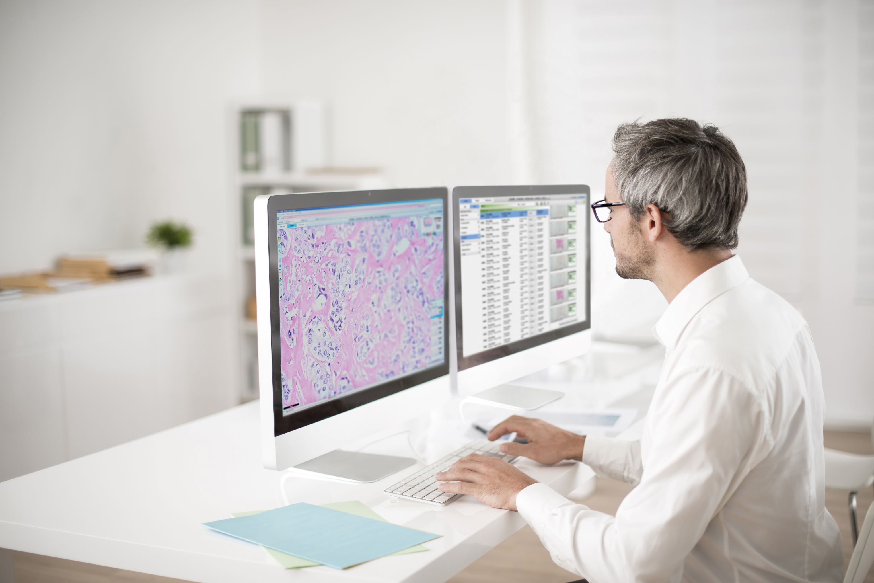
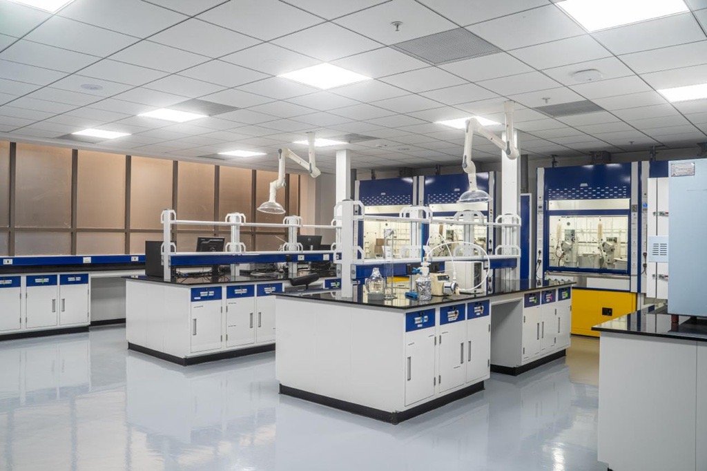
As part of the initiative supported by iHub-Data, IIIT-H installed a whole slide digital scanner at the premises of NIMS, Hyderabad. One of the first datasets that has been released is the IPD-Brain dataset in Nature Scientific Data – a prestigious, open-access, online-only journal for descriptions of scientifically valuable datasets.
With its focus on Indian demographics, it comprises 547 high-resolution H&E (haematoxylin and eosin stain) slides from 367 patients making it one of the largest in Asia.
While a beginning has been made by curating a dataset on brain tumours, efforts are underway to expand the dataset to include other cancers such as breast cancer, lung cancer, colorectal, oral and cervical cancers. NIMS is also contributing to curating the dataset on lung cancer.
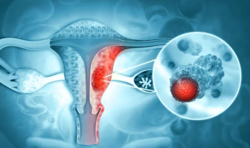
In addition to the cancer datasets, the project has also compiled another on lupus nephritis. Lupus is a kidney disease that occurs when the immune system attacks the kidneys.
While subtyping and grading of the cancers are routine and time-consuming tasks traditionally performed by histopathologists, tasks that cannot be performed by human observers are predicting molecular markers from H&E slide images. Molecular profile details are traditionally obtained from genetic labs where such testing is done or by performing immunohistochemistry (IHC). The group has instead attempted to predict molecular details with the help of tissue morphology itself using H&E slide images. One such effort involves predicting IDH mutations which plays a critical role in the diagnosis and prognosis of brain tumor patients.