26 May 2017 | News
The device provides higher resolution neural readings than existing tools used in the clinic and could enable doctors to perform safer, more precise brain surgeries.
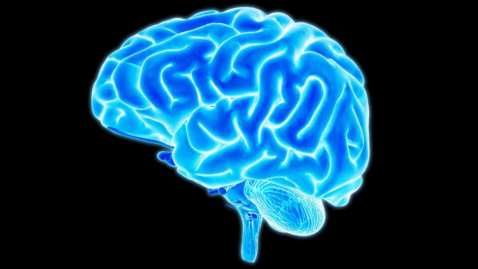
Researchers at the University of California, San Diego have developed a new device to map the brain during surgery and distinguish between healthy and diseased tissues. The device provides higher resolution neural readings than existing tools used in the clinic and could enable doctors to perform safer, more precise brain surgeries.
The device is an improved version of a clinical tool called an electrode grid, which is a plastic or silicone-based grid of electrodes that is placed directly on the surface of the brain during surgery to monitor the activity of large groups of neurons. Neurosurgeons use electrode grids to identify which areas of the brain are diseased in order to avoid damaging or removing healthy, functional tissue during operations. Despite their wide use, electrode grids have remained bulky and have not experienced any major advances over the last 20 years.
The new electrode grid is about a thousand times thinner than clinical electrode grids. This allows it to conform better to the intricately curved surface of the brain and obtain better readings.
The team's next steps are to make higher density electrode grids for improved resolution and biocompatibility tests to see how long they can stay in the body before they experience biofouling.