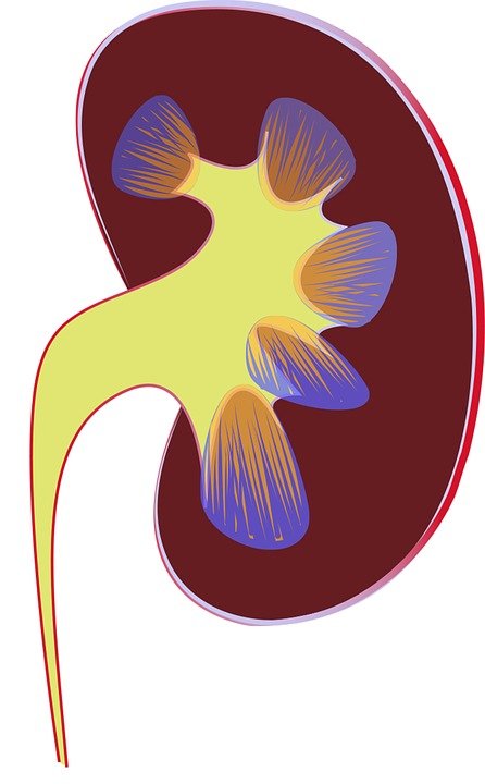
Optical spectroscopy for assessing kidney function
May 08, 2017 | Monday | News
It explores the use of multimodal auto fluorescence and light scattering to evaluate functional changes in the kidneys after ischemic injury.
A new technique developed by researchers at Lawrence Livermore National Lab promises to improve accuracy and lower costs of real-time assessment of kidney function.
It explores the use of multimodal auto fluorescence and light scattering to evaluate functional changes in the kidneys after ischemic injury. Conditions including accumulated arterial plaque or blood clots restrict the flow of oxygen and glucose to organs, and prolonged periods of such ischemia can compromise function.
While other current work in this area uses expensive multiphoton and laser-based techniques, the researchers reduced expenses by switching to camera-based imaging.
Currently, there is no real-time tool to measure the degree of ischemic injury incurred in tissue or to predict the return of its function. The inability to decisively determine tissue functional status runs two great risks: that dysfunctional tissue may be transplanted, increasing the morbidity and mortality of the patient; and that much-needed functional kidney tissue may be discarded.
Analysis of the light-scattering and autofluorescence images suggests that variations in tissue microstructure, fluorophore emission, and blood absorption spectral characteristics, combined with vascular response, contribute to the behavior of the recorded signals. These are used to obtain tissue functional information and enable the ability to predict post-transplant kidney function.