New microscopic technique to detect metastatic melanomas

05 May 2017 | News

This method could help medical doctors and pathologists to detect cancers as they spread, becoming one of the tools in the fight against this fatal disease.

Researchers at the University of Missouri Columbia have devised a new tool to detect and analyze single melanoma cells that are more representative of the skin cancers developed by most patients.

The team that included Gary Baker, an assistant professor of chemistry in the MU College of Arts and Science and Gerardo Gutierrez-Juarez, a professor and investigator at the University of Guanajuato in Mexico, decided to supplement an emerging technique called photoacoustic (PA) spectroscopy, a specialized optical technique that is used to probe tissues and cells non-invasively. Current systems use the formation of sound waves followed by the absorption of light which means that the tissues must adequately absorb the laser light. This is why, up until now, researchers have focused only on strong-light-absorb cells melanoma cells.

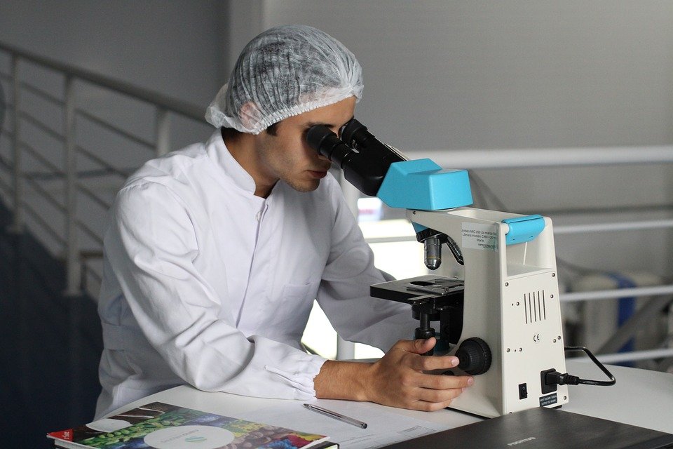
The team modified a microscope that was able to merge light sources at a range conducive to observing the details of single melanoma cells. Using the modified system, human melanoma and breast cancers as well as mouse melanoma cells were diagnosed with greater ease and efficiency. The team also noted that as the cancer cells divided, they grew paler in color but the system was able to detect the newer, smaller cells as well.

This method could help medical doctors and pathologists to detect cancers as they spread, becoming one of the tools in the fight against this fatal disease.

Comments

× Your session has been expired. Please click here to Sign-in or Sign-up
   New User? Create Account