“A good number of diseases can be diagnosed with a chest X-ray”
November 08, 2020 | Sunday | Interviews
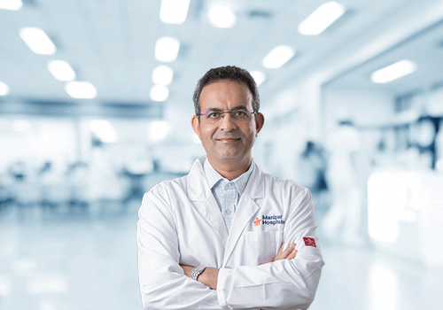
BioSpectrum spoke with Dr Sudarshan Rawat, HOD & Consultant – Radiology, Manipal Hospitals Old Airport Road, Bengaluru about the common challenges encountered by the radiologists during COVID and technological advancements in the area
As every year, the International Day of Radiology will be celebrated on November 8 with the aim of building greater awareness of the value that radiology contributes to safe patient care, and improving public understanding of the vital role radiologists and radiographers play in the healthcare continuum. This is a difficult time for the whole world as the spread of the COVID-19 pandemic affects so many areas of our lives. In challenging times like these, hope can be a powerful force for change and a compelling source of reassurance. The tireless and tremendous efforts and dedication of medical staff in fighting the pandemic gives everyone hope.
In 2020, the International Day of Radiology (IDoR2020) was dedicated to all imaging professionals and their essential role in fighting the COVID-19 pandemic making an indispensable contribution to the diagnosis and treatment of COVID-19 patients. BioSpectrum spoke with Dr Sudarshan Rawat, HOD & Consultant – Radiology, Manipal Hospitals Old Airport Road, Bengaluru, who is a national board examiner in Radiology, New Delhi and DNB teacher at Manipal Hospital, Bengaluru about the common challenges encountered by the radiologists during COVID and technological advancements in the area.
What are the common challenges encountered by the radiologists during COVID and what are being precautions taken by them for the same?
Number 1, the COVID 19 can affect several organs of the body, but it primarily affects the lungs and the blood vessels. Both the organs are best assessed by radiology namely a chest x-ray or CT for the lungs and a Doppler or angiography for the blood vessels. Many of these had to be repeated at intervals to assess the progress of the disease in infected patients.
Number 2, the healthcare professionals working in the Radiology Department such as Radiographers, Technicians and Radiologists are in close contact with the patient during these procedures. To avoid the spread of the virus, we use full protection (PPE, N 95 Masks, goggles/face shield) while handling COVID-19 patients and sanitize all equipment (x-ray-cassette, CT and USG machines) after a patients scan is done. All equipment and frequently touched surfaces are sanitized regularly on an hourly basis.
Because many infected patients are asymptomatic, we take all precautions when in close contact with any patient e.g. during ultrasound or biopsies and we ensure that proper hand sanitization is being practised in between handling the patients. We also try to keep minimal face-to-face contact with patients and discuss results etc over the phone or video consultations. Number 3, one of the most disturbing trends that we are witnessing is the late presentation of patients with cancers, strokes, heart attacks. It is very unfortunate that they delayed treatment and did not visit the hospital when they needed it the most.
The COVID-19 pandemic has created multiple challenges for medical imaging: the primary ones being diagnosing, monitoring the disease, follow up with or without related complications and most importantly the infection control among the staff, which was incredibly challenging. But we have been lucky enough to control the infection among the health care workers.
In the end, our message is Stay Safe and Stay Healthy too.
In the era of technological advancement does X-rays still play a role over CT and MRI?
Despite the advances, a simple x-ray is still a very important part of our diagnostic array. The chest X-ray is still the commonest radiological investigation requested. A good number of diseases can be diagnosed with a chest X-ray. A normal chest x-ray is very reassuring. X-rays are indispensable in assessing fractures and joints. X-rays are easily available, economical, and can be done at the bedside for sick patients. The radiation dose of an X-ray is minimal compared to other investigations.
-----------
“Wilhelm Conrad Röntgen produced the first X-ray image of his wife’s hand, which was the first medical imaging photo ever published in the first scientific article on medical imaging in December 1895, this breakthrough technology rapidly revolutionized medicine and earned Roentgen the first Nobel Prize in physics in 1901. It’s now 125 years since then, a new branch radiology was created, which has been evolved with digital x-rays, further invented ultrasound, digital mammogram, CT scan, PET-CT scan and MRI and changed the world in diagnosing the disease and cancers early, thus reducing disease related mortality and improving human life span, which was earlier 50-60 years and now 80-90 years, it is also one the most sought-after branch for specialization in medicine”.
Dr Sudarshan Rawat, HOD & Consultant – Radiology, Manipal Hospitals Old Airport Road, Bengaluru


